[TargetMol Star Molecule] - ML162: A Precise GPX4 Inhibitor Unleashing Ferroptosis to Probe Tumor Vulnerabilities
Read Time:12 Minute, 16 Second
■ 1. Background
ML162 (T8970), CAS: 1035072-16-2, is a covalent inhibitor of glutathione peroxidase 4 (GPX4) that potently induces ferroptosis, a regulated, iron-dependent cell death modality driven by unchecked phospholipid peroxidation [1,2]. Mechanistically, ML162 forms a covalent adduct with GPX4, a selenoenzyme that normally reduces phospholipid hydroperoxides to their corresponding alcohols using reduced glutathione (GSH) as an electron donor. By functionally inactivating GPX4, ML162 disables a central antioxidant checkpoint that restrains lipid radical propagation in polyunsaturated phospholipids, thereby promoting the accumulation of lipid hydroperoxides and secondary reactive species that execute ferroptotic demise of cells [1,2]. Within the ferroptosis pathway, GPX4 operates downstream of cystine import through system xC− (SLC7A11/SLC3A2), GSH synthesis, and NADPH supply, and in opposition to processes that enrich and oxidize polyunsaturated acyl chains in membranes—such as ACSL4-dependent phospholipid remodeling, enzymatic peroxidation by lipoxygenases, and iron-catalyzed Fenton chemistry. Because ML162 directly targets GPX4, its action bypasses upstream modulation of cystine transport and GSH availability; even in GSH-replete conditions, GPX4 inhibition by ML162 permits lipid peroxide accumulation to exceed the threshold for ferroptosis. In cellular contexts with abundant polyunsaturated phospholipid substrates or heightened labile iron, ML162-driven GPX4 blockade can be especially effective at triggering ferroptotic signaling.
Biologically, ML162 functions as a precise chemical probe to acutely extinguish GPX4 activity and thereby activate ferroptosis, enabling temporal dissection of the lipid peroxidation cascade and downstream cell death execution. Notably, ML162 exhibits selective growth inhibition of cell lines bearing mutant RAS oncogenes, aligning with early observations that RAS-driven states display ferroptosis vulnerabilities uncovered by small-molecule screening [1]. This selectivity creates a practical entry point to interrogate the metabolic and redox liabilities of RAS-mutant models, to stratify ferroptosis sensitivity phenotypes, and to evaluate how genetic or environmental modifiers (for example, variation in SLC7A11 expression or ACSL4-dependent lipid composition) intersect with GPX4 dependence [1,2].
As a pathway modulator, ML162 is suited for: benchmarking GPX4 dependence across panels of engineered or patient-derived cell lines; mapping ferroptosis-rescue networks using genetic perturbations; lipidomic profiling of oxidized phospholipid species as proximal ferroptotic effectors; and combination studies that compare GPX4 inhibition with interventions acting elsewhere in the pathway (such as limiting cystine supply or altering iron handling), thereby distinguishing upstream versus downstream ferroptosis control points. Reported antitumor activity of ML162 in experimental systems further supports its use to model ferroptosis-driven suppression of cellular proliferation and to explore contexts in which ferroptotic pressure constrains oncogenic fitness in vitro and in vivo-like settings [1,2]. Together, ML162 provides a robust, covalent handle on GPX4 to mechanistically activate ferroptosis and to probe the signaling architecture that couples lipid peroxide control to selective vulnerabilities in oncogene-defined states.
■ 2. Literature review
■ 2.1 The ferroptosis inducing compounds RSL3 and ML162 are not direct inhibitors of GPX4 but of TXNRD1
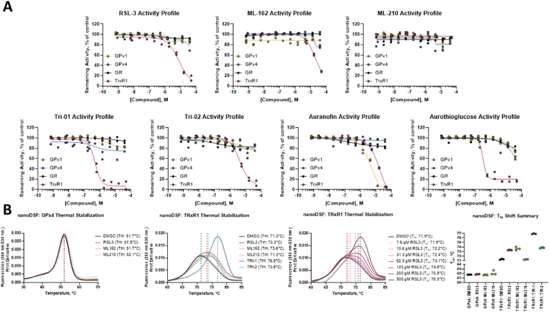
▲ In vitro pure enzyme activity assays show that class I FINs (RSL3, ML162, ML210) and known TXNRD1 inhibitors (TRi‑1, TRi‑2, Auranofin, Aurothioglucose) do not inhibit GPX1, GPX4, or GR, but effectively suppress TXNRD1 activity (A). NanoDSF thermal stability experiments further demonstrate that ML162 (100 μM), along with RSL3, TRi‑1, and TRi‑2, significantly stabilizes TXNRD1 protein, whereas ML210 does not (B). Data are presented as mean ± s.d. of n = 3 technical replicates.
ML162 (T8970) was identified as a highly efficient inhibitor of thioredoxin reductase 1 (TXNRD1). In pure enzyme assays, ML162 inhibited TXNRD1 with an IC 50 of 19.5 μM. In live A549 human lung cancer cells cultured with selenium supplementation and monitored using the TXNRD1-selective RX1 probe, ML162 produced a clear, dose-dependent suppression of cellular TXNRD1 activity at concentrations of 0.5 μM or higher. The onset of this suppression was rapid, with 4 hours of incubation at 1 μM or higher being sufficient to reduce the RX1 signal, and the cellular inhibition of TXNRD1 by ML162 showed similar efficacy to the benchmark inhibitor TRi-1. At later measurement timepoints under these culture conditions, exposure to ML162 resulted in significant cell death. Together, these findings demonstrate that ML162 directly inhibits TXNRD1 in vitro and suppresses TXNRD1 activity in cells in a dose-dependent and rapid manner, accompanied by cell death at later timepoints.[3]
■ 2.2 Targeting Wnt/β-Catenin Signaling Exacerbates Ferroptosis and Increases the Efficacy of Melanoma Immunotherapy via the Regulation of MITF
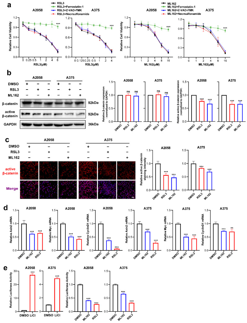
▲ In A2058 and A375 melanoma cells, ferroptosis inducers RSL3 and ML162 significantly reduce cell viability, an effect rescued by ferrostatin‑1 but not by Z‑VAD‑FMK or necrosulfonamide (a). ML162 (4 μM for both A2058 and A375) treatment for 24 h downregulates β‑catenin and active β‑catenin protein levels (b), reduces nuclear localization of active β‑catenin by immunofluorescence (c), suppresses mRNA expression of downstream targets Axin2, c‑Myc, and CyclinD1 (d), and inhibits β‑catenin transcriptional activity as shown by Topflash reporter assay (e). Data are mean ± SEM of triplicates; **p<0.01, ***p<0.001.
ML162 (T8970) is described as a potent small-molecule inducer of ferroptosis that targets GPX4 activity. In this study, ML162 was applied to melanoma cell lines to probe cellular responses relevant to ferroptosis and Wnt/β-catenin signaling. Cell viability was evaluated after exposure to ML162 across a 1–16 μM range for 24 hours, with parallel pretreatments using ferrostatin-1, Z-VAD-FMK, or necrosulfonamide to contextualize the type of cell death. To assess signaling consequences, A2058 and A375 melanoma cells were treated with 4 μM ML162 for 24 hours, followed by immunoblotting of β-catenin and active β-catenin, as well as immunofluorescence staining of active β-catenin. The study further quantified the relative mRNA levels of canonical Wnt/β-catenin targets Axin2, c-Myc, and CyclinD1 after ML162 treatment. Complementing these measurements, β-catenin transcriptional activity was examined via a Topflash reporter assay in both A2058 and A375 cells following 24-hour exposure to 4 μM ML162. Together, these ML162-directed experiments position the compound as a GPX4-targeting ferroptosis inducer and as a tool to interrogate β-catenin signaling and transcriptional readouts in melanoma cells.[4]
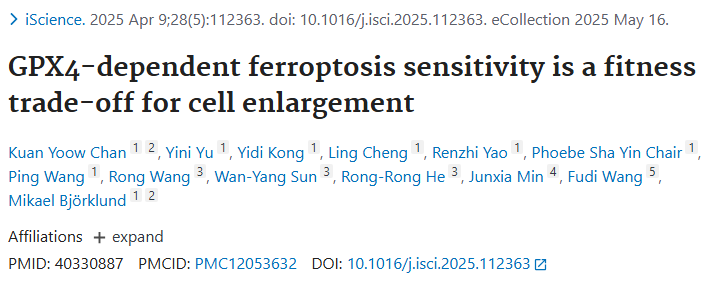
■ 2.3 GPX4-dependent ferroptosis sensitivity is a fitness trade-off for cell enlargement
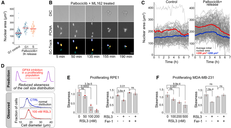
▲ In RPE1‑mScarlet‑PCNA cells, palbociclib (200 nM, 48 h) pretreatment followed by release synchronizes cell cycle and alters nuclear area distribution in G1 and S phase cells (A). SC‑Track tracing shows dynamic nuclear area changes upon palbociclib plus ML162 treatment (B). Single‑cell nuclear area trajectory analysis reveals that ML162 preferentially eliminates large cells (nuclear area >200 μm²) in the palbociclib‑pretreated group, whereas control cells and those <200 μm² are less affected (C). The observed effect of RSL3 on cell size distribution in proliferating populations matches theoretical predictions (D), and RSL3 dose‑dependently alters skewness in RPE1 (E) and MDA‑MB‑231 (F) populations, an effect rescued by Fer‑1. Data are mean ± SD, n=3; ANOVA with Tukey’s post‑hoc test.
ML162 (T8970) is identified in this study as a GPX4 inhibitor. The authors report that large cells show a marked potentiation of response to ML162, with more than a 100-fold increase in sensitivity observed under enlarged-cell conditions. This places ML162 among compounds whose activity is strongly amplified when cell size increases, directly linking the magnitude of cellular response to the state of cellular enlargement. The evidence explicitly characterizes ML162 within the GPX4 inhibitor class and quantifies the sensitivity shift as exceeding 100-fold in large cells. Together, these findings define a pronounced size-dependent effect for ML162 in this experimental system, highlighting that increased cell size strongly augments the compound’s measured activity.[5]
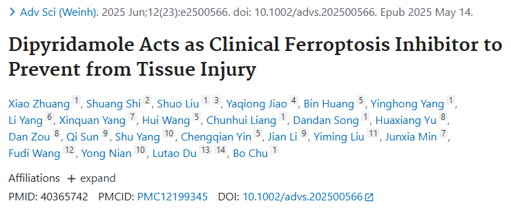
■ 2.4 Dipyridamole Acts as Clinical Ferroptosis Inhibitor to Prevent from Tissue Injury
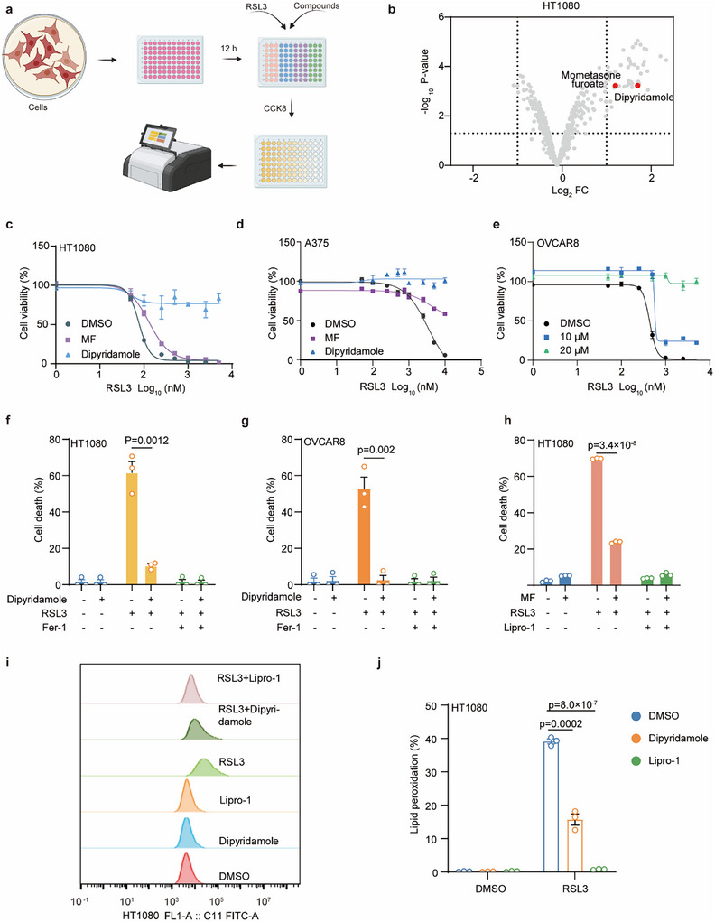
▲ A clinical drug screen was performed in HT1080 cells to identify candidate ferroptosis inhibitors (a). Volcano plot highlights dipyridamole and MF (mometasone furoate) as top protective hits (b). Dipyridamole or MF pretreatment dose‑dependently reduces RSL3‑induced cell death in HT1080 (c), A375 (d), and OVCAR8 (e) cells. SYTOX Green staining confirms that dipyridamole, Fer‑1, and Lipro‑1 significantly reduce RSL3‑induced cell death in HT1080 and OVCAR8 cells (f–h). BODIPY 581/591 C11 staining shows that dipyridamole and Lipro‑1 suppress RSL3‑induced lipid peroxidation (i, j). Data are mean ± SEM, n=3 biologically independent experiments; two‑tailed unpaired Student's t‑test.
ML162 (T8970) was employed in this study as one of several alternative methods to provoke ferroptotic cell death, alongside cystine/cysteine starvation, erastin, and ML210. The paper explicitly states that ferroptotic cell death was induced using ML162, indicating its functional role in triggering this regulated form of cell death within the experimental framework. ML162’s inclusion in this panel of inducers was part of the approach used to establish ferroptosis in cellular systems for subsequent analyses, which are referenced in the Supporting Information figures. By utilizing ML162 to reliably elicit ferroptotic cell death, the investigators incorporated it into their comparative experiments designed to assess cellular responses under ferroptotic stress. The text clearly situates ML162 among the agents capable of initiating ferroptotic death in the models examined, thereby defining its role in the study as an inducer used to establish the ferroptosis context necessary for downstream evaluations. The supporting materials cited for these inducer-based experiments include Figure S1e–h, which correspond to the assays where alternative ferroptosis-inducing conditions, including ML162, were applied.[6]
■ 2.5 METTL17 coordinates ferroptosis and tumorigenesis by regulating mitochondrial translation in colorectal cancer
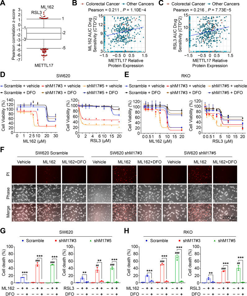
▲ METTL17 expression positively correlates with cancer cell resistance to GPX4 inhibitors (ML162 and RSL3) (A). DepMap database analysis reveals significant positive correlations between ML162 (B) or RSL3 (C) drug sensitivity (AUC) and METTL17 protein levels in colorectal and other cancer cell lines. Knockdown of METTL17 (shM17#3, #5) significantly enhances ML162‑ or RSL3‑induced viability reduction in SW620 (D) and RKO (E) cells, an effect partially rescued by the iron chelator DFO. Microscopy with PI staining confirms that ML162‑induced cell death is more pronounced in METTL17‑knockdown cells (F). Quantification shows that METTL17 knockdown significantly increases PI‑positive cell death upon ML162 or RSL3 treatment, which is reversed by DFO (G, H). Data are mean ± SD, n=3–4; two‑tailed Student's t‑test or Pearson correlation test. ##p<0.01, ###p<0.001 vs. ML162/RSL3‑treated Scramble; **p<0.01, ***p<0.001 vs. indicated groups.
ML162 (T8970) was used to probe ferroptosis in colorectal cancer models. The study explicitly describes ML162-induced ferroptosis, as restoring METTL17 expression in knockdown SW620 cells partially rescued ML162-induced ferroptosis, while METTL17 overexpression in control cells slightly promoted this response. METTL17 depletion increased sensitivity to ML162 across systems: SW620 xenografts were sensitized to ML162 with reductions in tumor volume, size, and weight, and knockdown of METTL17-interacting proteins LRPPRC, MRPS9, MRPS22, and MRPS35 sensitized RKO cells to ML162-induced ferroptosis. Mechanistically, ML162 exposure elevated oxidative and mitochondrial stress: after 3 hours of ML162 treatment, SW620 cells displayed increased lipid peroxides, and in METTL17-deficient cells ML162 further increased intracellular and mitochondrial ROS and was associated with pronounced mitochondrial damage, including mitochondrial shrinkage and loss of cristae. Bioenergetically, in SW620 cells with METTL17 knockdown, ML162 treatment decreased cellular NADH. ML162 also coincided with protein-level changes in ferroptosis-related markers under METTL17 deficiency, with suppression of HO-1 and upregulation of NRF2 after ML162 exposure. At the systems level, expression profiling across datasets linked higher levels of LRPPRC, MRPS9, MRPS22, and MRPS35 with resistance to ML162, consistent with the observed sensitization to ML162-induced ferroptosis upon their knockdown.[7]
■ 3. Conclusion
ML162 functions as a covalent inhibitor of glutathione peroxidase 4 (GPX4), blocking its ability to reduce phospholipid hydroperoxides and thereby triggering ferroptosis, an iron-dependent form of regulated cell death. Literature reports emphasize ML162’s role in acutely extinguishing GPX4 activity to induce lipid peroxidation and ferroptotic cell death, with heightened sensitivity in RAS-mutant and enlarged cells. Additionally, ML162 inhibits TXNRD1 at higher concentrations, contributing to cellular oxidative stress and death. Studies demonstrate its utility in probing ferroptosis signaling pathways, including Wnt/β-catenin regulation in melanoma and mitochondrial translation control via METTL17 in colorectal cancer, highlighting its versatility as a chemical probe. Future research may focus on leveraging ML162 to map ferroptosis-rescue networks, explore metabolic vulnerabilities in diverse oncogenic contexts, and develop combination therapies that exploit ferroptotic pressure to suppress tumor growth.
■ 4. Q&A
Q1: What is the primary mechanism by which ML162 induces ferroptosis?
A1: ML162 covalently inhibits GPX4, preventing it from reducing phospholipid hydroperoxides, leading to accumulation of lipid peroxides and triggering iron-dependent ferroptotic cell death.
Q2: How does ML162’s activity relate to cell size and genetic background?
A2: ML162 sensitivity is markedly increased in enlarged cells (over 100-fold) and shows selective growth inhibition in RAS-mutant cell lines, indicating that both cell size and oncogenic mutations influence ferroptosis susceptibility mediated by ML162.
Q3: Besides GPX4, does ML162 affect other cellular targets?
A3: Yes, ML162 also inhibits thioredoxin reductase 1 (TXNRD1) in a dose-dependent manner at micromolar concentrations, contributing to oxidative stress and subsequent cell death.
■ 5. Reference
[1] Dixon SJ, Lemberg KM, et al. Ferroptosis: An iron-dependent form of nonapoptotic cell death. Cell. 2012;149(5):1060-1072.
[2] Yang WS, SriRamaratnam R, et al. Regulation of ferroptotic cancer cell death by GPX4. eLife. 2014;3:e02523.
[3] Cheff D, Huang C, Scholzen K, Gencheva R, Ronzetti M, Cheng Q, et al.. The ferroptosis inducing compounds RSL3 and ML162 are not direct inhibitors of GPX4 but of TXNRD1. Redox Biology. 2023;62():102703.
[4] Wang H, Zhang H, Chen Y, Wang H, Tian Y, Yi X, et al.. Targeting Wnt/β-Catenin Signaling Exacerbates Ferroptosis and Increases the Efficacy of Melanoma Immunotherapy via the Regulation of MITF. Cells. 2022;11(22):3580.
[5] Chan K, Yu Y, Kong Y, Cheng L, Yao R, Yin Chair P, et al.. GPX4-dependent ferroptosis sensitivity is a fitness trade-off for cell enlargement. iScience. 2025;28(5):112363.
[6] Zhuang X, Shi S, Liu S, Jiao Y, Huang B, Yang Y, et al.. Dipyridamole Acts as Clinical Ferroptosis Inhibitor to Prevent from Tissue Injury. Advanced Science. 2025;12(23):.
[7] Li H, Yu K, Hu H, Zhang X, Zeng S, Li J, et al.. METTL17 coordinates ferroptosis and tumorigenesis by regulating mitochondrial translation in colorectal cancer. Redox Biology. 2024;71():103087.
www.targetmol.com
TargetMol Chemicals Inc.


Happy
0 %

Sad
0 %

Excited
0 %

Sleepy
0 %

Angry
0 %

Surprise
0 %
 0
0
 0
0


Average Rating